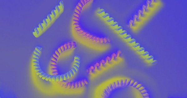
Researchers at MIT and Beihang University have developed a coiled nanofiber yarn that keeps living cells viable even when stretched to several times its original length, a breakthrough that could let patients move freely while muscle and tendon injuries heal. The innovation, described in a paper published Wednesday in the Proceedings of the National Academy of Sciences, may one day replace the restrictive boots and casts that currently immobilize patients during recovery.
"When you repair muscle or tendon, you really have to fix their movement for a period of time, by wearing a boot, for example," MIT research scientist Ming Guo told MIT News. "With this nanofiber yarn, the hope is, you won't have to [be] wearing anything like that."
The team engineered the yarn by twisting hundreds of thousands of aligned nanofibers, made from biocompatible materials, into tight coils. They then seeded different types of live cells—including muscle cells—along the coil structure. When the coils were stretched to six times their original length, most cells survived and continued to grow, according to the researchers. The coil shape appeared to "shelter" the cells from the mechanical forces that would normally kill them during movement, Guo explained in an interview with the MIT blog.
Why the Coil Design Matters
The key to the technique lies in the geometry of the coils. By mimicking the natural helical structure of muscle and tendon tissue, the yarn distributes strain away from the cells, preventing the membrane damage that typically occurs when cells are stretched. This protective effect allows the cells to remain viable and proliferate even under significant mechanical loading, a critical requirement for functional tissue repair.
Guo and his colleagues believe the approach could be adapted to a wide range of materials and cell types. "We may be able to one day embed these structures under the skin, and the [coil] material would eventually be digested, while the new cells stay put," he told MIT News. "The nice thing about this method is, it's really general, and we can try different materials. This may push the limit of tissue engineering a lot."
Implications for Recovery
Current treatments for severe muscle and tendon injuries often require weeks or months of immobilization, which can lead to muscle atrophy, joint stiffness, and prolonged rehabilitation. If the nanofiber yarn proves effective in clinical settings, it could allow patients to maintain mobility and muscle function during the healing process, potentially speeding up recovery and improving outcomes.
The research is still at an early stage, with tests conducted in laboratory settings rather than in living organisms. The team plans to explore different biocompatible materials and further refine the coil structure to optimize cell survival and integration with host tissue.
While the concept is promising, experts caution that significant hurdles remain before the technology can be translated into clinical practice. These include ensuring long-term stability of the yarn in the body, preventing immune rejection, and demonstrating efficacy in animal models and human trials.
Nevertheless, the study adds to a growing body of work on advanced tissue engineering, including efforts to 3D-print body parts for athletes and other patients. The MIT and Beihang collaboration highlights the potential of biomimetic designs to solve some of the most challenging problems in regenerative medicine.















Comments (0)